Abstract
Introduction: Preeclampsia and eclampsia area leading cause of maternal and fetal mortality with vasospasm and endothelial dysfunction as underlying pathophysiology. Considering the high incidence of maternal and fetal complications, obstetricians are confronted to identify the clinical feature or any parameter, which would alarm them of the high-risk to the mother or the fetus. The various pathological changes in different organs in the body can be studied directly by viewing the ocular fundus, which may act as a window to systemic vascular system. Objectives: Our aim was to determine the prevalence of fundus changes and its relevance to maternal and fetal outcome in patients of preeclampsia and eclampsia. Study design: Cross-sectional analytical study. Material and methods: A total of 150 pregnant mothers with preeclampsia and eclampsia were recruited in the study. They were evaluated for hypertensive retinopathy by direct ophthalmoscope and classified according to Keith-Wagener’s classification. Maternal outcomes were assessed in terms of complications, mode of termination of delivery and maternal mortality. Fetal outcomes were noted in terms of birth weight, Apgar score at birth, need for neonatal intensive care unit (NICU) admissions and perinatal mortality. Data obtained was subjected to statistical analysis using SPSS software and chi-square and student t-test was applied for the test of significance. Results: Hypertensive retinopathy was observed in 46% of patients. Statistically significant association of fundus changes were seen with severity of hypertension, mode of termination of pregnancy, maternal complications and perinatal outcome. Conclusions: Fundoscopy is a safe, simple, noninvasive and cost-effective procedure which can be used as a valuable tool to assess severity of preeclampsia and eclampsia and predicting adverse pregnancy outcome.
Keywords: Preeclampsia, hypertensive retinopathy, fundoscopy
Preeclampsia and eclampsia syndrome is a specific pregnancy disorder which occurs in late pregnancy and has potentially devastating consequences for both mother and baby. Together they form one member of the deadly triad along with hemorrhage and infection that contribute greatly to maternal morbidity and mortality. It causes myriad complications and ultimately results in multiorgan involvement with a clinical spectrum ranging from barely noticeable to life-threatening cataclysmic pathophysiological deterioration.1
Preeclamptic and eclamptic disorders also affect visual pathways, from the anterior segment to visual cortex with their potential impact on ocular fundus affecting 30% to 100% cases.2 The underlying pathophysiology is endothelial dysfunction and vasospasm of vessels in the retina similar to that, which occurs elsewhere in the body.3 Retinal angiospasm is an almost constant accompaniment of toxemia of pregnancy. When marked spastic changes occur early in pregnancy, they indicate presence of severe toxemic process which, unless controlled or pregnancy terminated, endangers the viability of the fetus and life of mother.4 Recent epidemiological studies have indicated that retinal vasculature reflects changes in systemic vasculature and shares anatomical and physiological similarities with microvasculature in other major organs such as brain, heart and kidney.5 The retinal blood vessels of 100 to 300 µm in size can be visualized noninvasively by fundoscopy which is a simple and cost-effective procedure. The ocular fundus has proved to be valuable and a valid prognostic procedure for assessment of severity of preeclampsia and neonatal outcome.6 Hence, the present study was designed with a view to find out prevalence and spectrum of fundus changes in patients with preeclampsia and eclampsia, association of fundus changes with fetomaternal outcome and various clinical and laboratory manifestations.
Material and Methods
This study was conducted in the Dept. of Obstetrics and Gynecology, LLRM Medical College and associated SVBP Hospital, Meerut from November 2013 to May 2015. A total number of 150 cases of preeclampsia and eclampsia were enrolled in the study.
Inclusion Criteria
- Age group 18-38 years.
- Women with ≥20 weeks of gestation diagnosed as preeclampsia and eclampsia.
- Cases with singleton or multiple pregnancy.
- All primigravida and multigravida fitting into the criteria of inclusion.
Exclusion Criteria
- Cases with known case of essential hypertension.
- Patients with diabetes/gestational diabetes.
- Patients with cardiovascular disease.
- Patients of collagen vascular disease.
- Patients with ocular media opacity (cataract, corneal opacity).
- Patients with chronic renal failure/disease.
Detailed history of women including present and past pregnancies, duration of gestation, any associated complaint, past history of any medical illness, epilepsy, chronic hypertension, diabetes, cardiovascular and collagen vascular disease was taken. A thorough general and obstetric examination was done and routine and specific investigations were undertaken. Antihypertensive drugs, magnesium sulfate regime and further management was tailored as per requirement of individual patient.
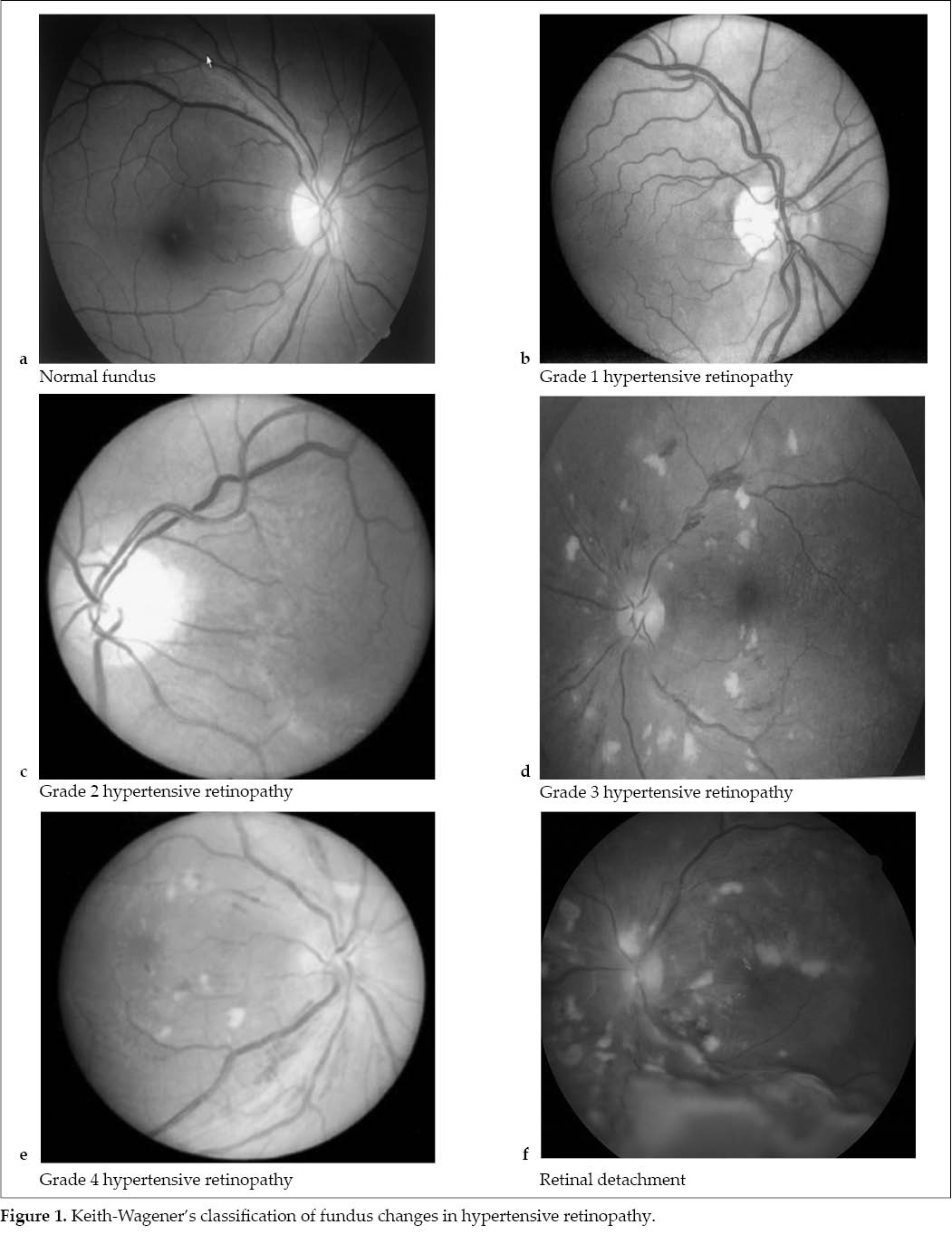
Ocular evaluation was done which included bedside visual acuity and torch light examination to rule out any gross anterior segment pathology and to elicit pupillary reflex. Fundus examination was done using direct ophthalmoscope after dilating the pupils with one drop of plain tropicamide (1%) instilled in each eye thrice at 10 minutes interval. Hypertensive retinopathy changes were graded according to Keith-Wagener’s classification (Fig. 1 a-f). Maternal outcome were recorded in relation to fundus changes, mode of delivery and complications. Fetal outcomes were noted in terms of weight, age, Apgar score, neonatal intensive care unit (NICU) admissions and mortality. The data obtained was subjected to statistical analysis. P <0.05 indicated statistically significant and <0.01 was considered highly significant.
Results
A total number of cases were studied over a period of 1½ year. The study group comprised of 49.3% cases with nonsevere preeclampsia, 27.3% with severe preeclampsia and 23.3% with eclampsia. The mean age of the patients in our study was 25.87 years (standard deviation [SD] - 4.19 years) and the mean gestational age at the time of presentation was 37 weeks (SD 3 weeks). Majority of the cases were primigravidas (52%). Among our subjects most common symptom observed was swelling on legs and face in 89.3% of cases. Headache was the next common symptom (40.7%) followed by epigastric pain (32%), blurring of vision (30%), convulsions (23.3%) and complete reversible loss of vision was reported in one patient.
Hypertensive retinopathy was observed in 46% of cases, 24% had Grade 1 changes. Grade 2 and Grade 3 changes were seen in 16% and 4.7%, respectively. Grade 4 changes were seen in 1.3% of cases and 2 women had retinal detachment (Table 1).
Mean systolic and diastolic blood pressure in cases without fundus changes was 152.62 ± 11.71 mmHg and 97.68 ± 7.54 mmHg, respectively and in cases with fundus changes, it was 170.93 ± 14.53 mmHg and 105.71 ± 10.97 mmHg, respectively. The difference was significant with p value <0.05 (Table 2).
In our study, cases having 2+ proteinuria or more showed increased incidence of fundus changes (p <0.001). It was seen that 76.8% cases with fundus changes had deranged liver function tests compared to 22.2% in women with normal fundus (p <0.001). Similarly, we observed higher incidence of deranged renal function in women with hypertensive retinopathy (39.1%) than women who had normal fundus (2.5%; p <0.001). Also, 30.4% cases with fundus changes had decreased platelet count compared to only 3.7% in those without fundus changes (p <0.05). These results were statistically highly significant (Table 3).
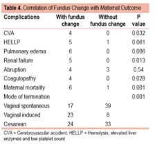
Higher incidence of maternal complications were seen in women with fundus changes. Statistically significant association was found between mode of termination of pregnancy and fundus changes (p = 0.047). More number of women with fundus changes were induced for obstetric reasons and uncontrolled blood pressure. Incidence of maternal mortality was also significantly higher in cases with hypertensive retinopathy with p <0.05 (Table 4).
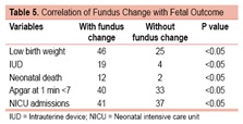
Fetal outcomes were assessed in all the cases as shown in Table 5. Cases with fundus changes had higher incidence of low birth weight babies (p <0.05), intrauterine deaths (p <0.05), early neonatal deaths (p <0.05), Apgar <7 at 1 minute and NICU admissions (p <0.05).
Discussion
Mean age of women in our study was 25.8 years. This was in consistence with other studies done by Jaeffe and Schatz,1 Tadin et al,2 Karki et al3 and Reddy et al.4
Majority that is 52% of cases were primigravidas. Similar were the observation by, Shah et al5 (50.7%). Higher percentages were observed by Doddamani et al6 (65.3%) and Reddy et al4 (65.3%).
Mean gestational age was 37 weeks in our study. This was in accordance to studies by Tadin et al2 (36.1 weeks), Saxena et al7 (37.4) and Nawaz et al8 (37.3).
Retinal changes have been observed in 30% to 100% of patients with preeclampsia and eclampsia according to literature. In the present study, 46% of subjects had hypertensive retinopathy. The prevalence rate was similar when compared to studies like Tadin et al2 (45%), Revathy9 et al (50%) and Bhandari et al10 (44%). On the contrary, lower prevalence was reported by
Karki et al3 (13.7%) and Rasdi et al11 (21.5%). Further, Grade 1 hypertensive retinopathy which consists of retinal arterial narrowing was the most common grade of retinopathy, seen in 24% women. The results are similar to the studies of Rasdi et al,11 Reddy et al,4 Bhandari et al10 and Bharathi12 et al. Retinal detachment was seen in 1.3% subjects in our study, which was comparable to a recent study by Bharathi et al.12
Mean systolic and diastolic blood pressure for subjects without fundus change were 152 mmHg and 96 mmHg, respectively and for subjects with fundus changes were 170 mmHg and 106 mmHg, respectively. Our result is supported by Reddy et al,4 Bhandari et al,10 Karki et al3 and others except for the study done by Rasdi et al.11
Our study found significant association between severity of proteinuria and severity of hypertensive retinopathy (p <0.05). This is supported by the studies conducted by Revathy et al,9 Shah et al,5 Mithila et al13 and Reddy et al.4
We found a statistically significant association between mode of termination of pregnancy and presence of fundus changes which is in accordance with study by Revathy et al.9 In contradiction Javadekar et al,14 and Ranjan et al15 did not find a significant association.
Hypertensive retinopathy has long been known as a predictor of systemic morbidity and mortality. In our study, significant positive association was found between fundus changes, maternal complications and maternal mortality, which is supported studies by Revathy et al,9 Ranjan et al15 and Rajalaxmi et al.16
Some studies reported poor fetal prognosis while others reported no prognostic implications on the fetus. Our study found a significant correlation between fundus changes and various perinatal outcome like prematurity, low birth weight, intrauterine death, neonatal death, Apgar score and NICU admissions (p <0.05). Javadekar et al,14 found a significant association between fundus changes, prematurity and low birth weight. Mithila et al,13 found a significant association between birth weight and hypertensive retinopathy. Doddamani et al,6 observed higher incidence of low birth weight, perinatal mortality and NICU admissions in women with preeclampsia and eclampsia having hypertensive changes in their fundus. Our study contradicts the study of Karki et al,3 who observed no significant association between presence of fundus changes with fetal outcome in terms of Apgar score at 1 minute, neonatal death and stillbirth. Ranjan et al15 also did not find significant association between fundus changes and Apgar score (p >0.05).
Conclusion
The study implies that degree of hypertensive retinopathy in women with preeclampsia and eclampsia is a valid and reliable parameter that gives prognostic information in assessment of severity of preeclampsia and neonatal outcome. Ophthalmoscopy is a simple, bedside, cost-effective and noninvasive procedure serving as a window to systemic vasculature and hence helping the obstetrician in assessing the severity of disease and guiding subsequent obstetric management of the patient. Timely ophthalmoscopy should be called for in all cases of pregnancy-induced hypertension as it would affect the decision of termination, thereby preventing other complications.
Acknowledgment
We are extremely thankful to all the women who participated in the study and the Dept. of Ophthalmology for their kind cooperation.
References
- Jaffe G, Schatz H. Ocular manifestations of preeclampsia. Am J Ophthalmol. 1987;103(3 Pt 1):309-15.
- Tadin I, Bojic L, Mimica M, Karelovic D, Dogas Z. Hypertensive retinopathy and pre-eclampsia. Coll Antropol. 2001;25 Suppl:77-81.
- Karki P, Malla P, Das H, Uprety DK. Association between pregnancy-induced hypertensive fundus changes and fetal outcomes. Nepal J Ophthalmol. 2010;2(1):26-30.
- Reddy SC, Nalliah S, George SR a/pKovil, Who TS. Fundus changes in pregnancy induced hypertension. Int J Ophthalmol. 2012;5(6):694-7.
- Shah AP, Lune AA, Magdum RM, Deshpande H, Shah AP, Bhavsar D. Retinal changes in pregnancy-induced hypertension. Med J DY Patil Univ. 2015;8(3):304-7.
- Doddamani UG, Doddamani GB. Perinatal outcome in pre-eclampsia: a prospective study. Sch J Appl Med Sci. 2014;2(1C):291-3.
- Saxena S, Srivastava PC, Thimmaraju KV, Mallick AK, Dalmia K, Das B. Socio-demographic profile of pregnancy induced hypertension in a tertiary care centre. Sch J Appl Med Sci. 2014;2(6D):3081-6.
- Nawaz F, Sultan S, Siddiqui I. Pregnancy outcome in primigravida complicated with pregnancy induced hypertension. J Med Sci. 2014;22(1):46-8.
- Revathy K, Varalakhshmi B. Fundoscopic changes in PIH and association with maternal and fetal outcomes. Indian J Appl Res. 2015;5(9):473-4.
- Bhandari AJ, Bangal SV, Gogri PY. Ocular fundus changes in pre-eclampsia and eclampsia in a rural set-up. J Clin Ophthalmol Res. 2015;3:139-42.
- Rasdi AR, Nik-Ahmad-Zuky NL, Bakiah S, Shatriah I.Hypertensive retinopathy and visual outcome in hypertensive disorders in pregnancy. Med J Malaysia. 2011;66(1):42-7.
- Bharathi NR, Raju NRS, Prasad PK, Raju RS, Premalatha, Mayee K, et al. Fundus changes in pregnancy induced hypertension: a clinical study. J Evolut Med Dent Sci. 2015;4(09):1552-62.
- Mithila R, Narendra PD, Gomathy E, Krishnamurthy D. Study of association of fundal changes and fetal outcomes in preeclampsia. J Evolut Med Dent Res. 2014;3(21):5894-901.
- Javadekar SD, Javadekar DP, Joshi K, Khatiwala R. Fundoscopic changes in pregnant mother with hypertension complicating pregnancy and various parameters of foetus. Int J Recent Trends Sci Technol. 2013;7(3):110-3.
- Ranjan R, Sinha S, Seth S. Fundus changes and fetal outcomes in pregnancy induced hypertension: an observational study. Int J Sci Study. 2014;2(7):6-9.
- Rajalaxmi KK, Nayak SR. Preeclampsia/eclampsia and retinal micro vascular characteristics affecting maternal and foetal outcome: A prospective study amongst South Indian pregnant women. Int J Innovat Res Develop. 2013;2(11):444-8.